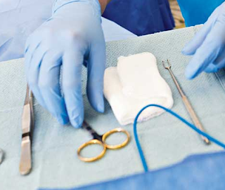
A specialized intravenous (IV) line known as a Broviac central venous line is placed into a large vein that travels from the chest wall to the heart. In cases where a young patient may require prolonged IV therapy, this solution is employed. Broviacs are available with one or two lumens. In general, doctors recommend using the smallest possible number of lumens, and experts only prefer using double-lumen catheters where there is an indication that calls for two infusions to be given at the same time. This is because venous thrombosis is more likely to occur in double-lumen catheters due to their bigger diameter. Additionally, a higher risk of infection is linked to an increase in catheter lumens.
Broviac Catheter Size
Both the Broviac and the Hickman catheters have a length of 90 centimeters, are radiopaque, and feature a Dacron cuff that is positioned 30 centimeters away from the Luer lock at the external end. With a single lumen, Broviac catheters are currently manufactured in sizes ranging from 2.7F (0.5 mm ID) to 7F (0.8 and 1.0 mm ID) with two lumens. The extravascular segment of Hickman catheters is covered with one or two Dacron cuffs and has one, two, or three lumens.

Broviac Catheter Insertion
Broviac catheters are typically inserted into patients while they are under the influence of anesthesia. In order to gain access to the jugular vein, the surgeon will need to make an incision. After that, the catheter is inserted further into the jugular vein until it meets the superior vena cava, which is the vein that leads directly into the heart. This enables doctors to effectively provide medications straight into the core of the circulatory system, a process known as "central vein access."
Broviac Catheter Placement
For the placement of the catheter, the jugular, subclavian, or femoral vein may be chosen as the most suited location. As needed, ultrasonography is used to determine the patency of this vein. The region is subsequently prepared and covered in sterile clothing as per the conventional surgical procedure. Placing a broviac line typically requires local anesthesia, either alone or in combination with intravenous sedation. Under fluoroscopy, the vein is accessed and a guidewire is inserted into the heart and then the inferior vena cava to validate the venous access point. At this point, the catheter exit site is chosen and a minor incision is performed. It is located about 2 fingerbreadths inferior to the venous puncture site. The catheter is then tunneled bluntly beneath the skin from the chest incision/exit site to the venotomy site, where it is inserted into the vein via a peel-away covering, which is then withdrawn. In order to prevent catheter-related thrombosis, the broviac catheter is cleansed postoperatively with heparin or alcohol/saline in patients who are sensitive to heparin.
Broviac Catheter Removal
Due to the fact that the dacron cuff expands into the subcutaneous tissues, its removal necessitates the utilization of an aseptic method and local anesthesia, in addition to the sharp cutting of the cuff from the subcutaneous tissues. After this, a sterile bandage is put on the catheter exit point on the chest, and pressure is maintained. In most cases, it takes a few weeks for the tract and the location where the catheter exited the body to heal.
Broviac Catheter Care
Constant looping and securing of the catheter are crucial. When working with smaller Broviacs, it is imperative that every inch of the thinner tubing be fastened firmly under the dressing. Heparin is a medication that is given to patients in order to avoid blood from clotting while the catheter is in place. Heparin must be used to flush the catheter once daily and after each use.


0 Comments
For comments please reply here.......